Written with StackEdit.
Gallvägar
Pre: Fastande, så att gallblåsan är dilaterad med anechoic galla.
Per: Liggande vänster sida, då kommer gallblåsan mer i mittlinjen.
-
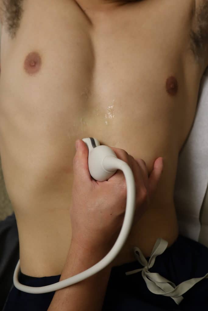
epigastriet, long-axis (Sagittal, S)
-
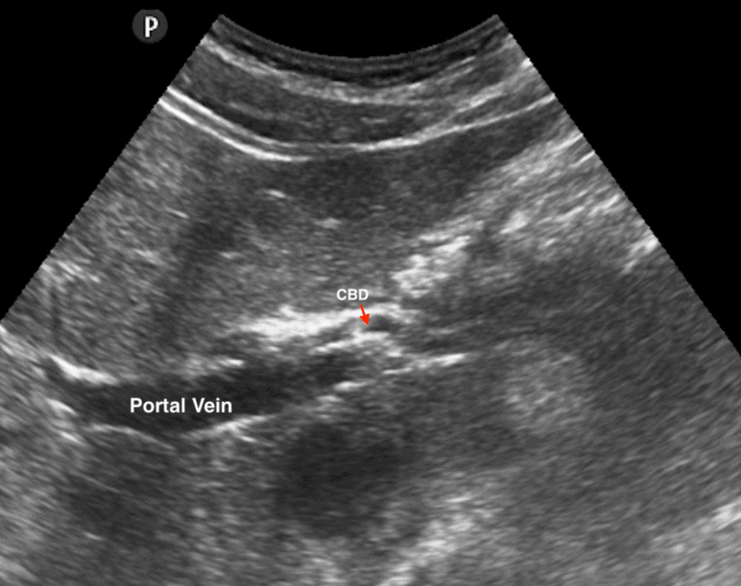
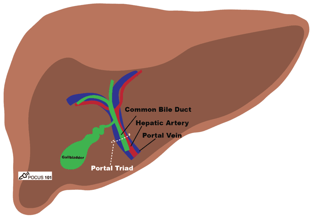
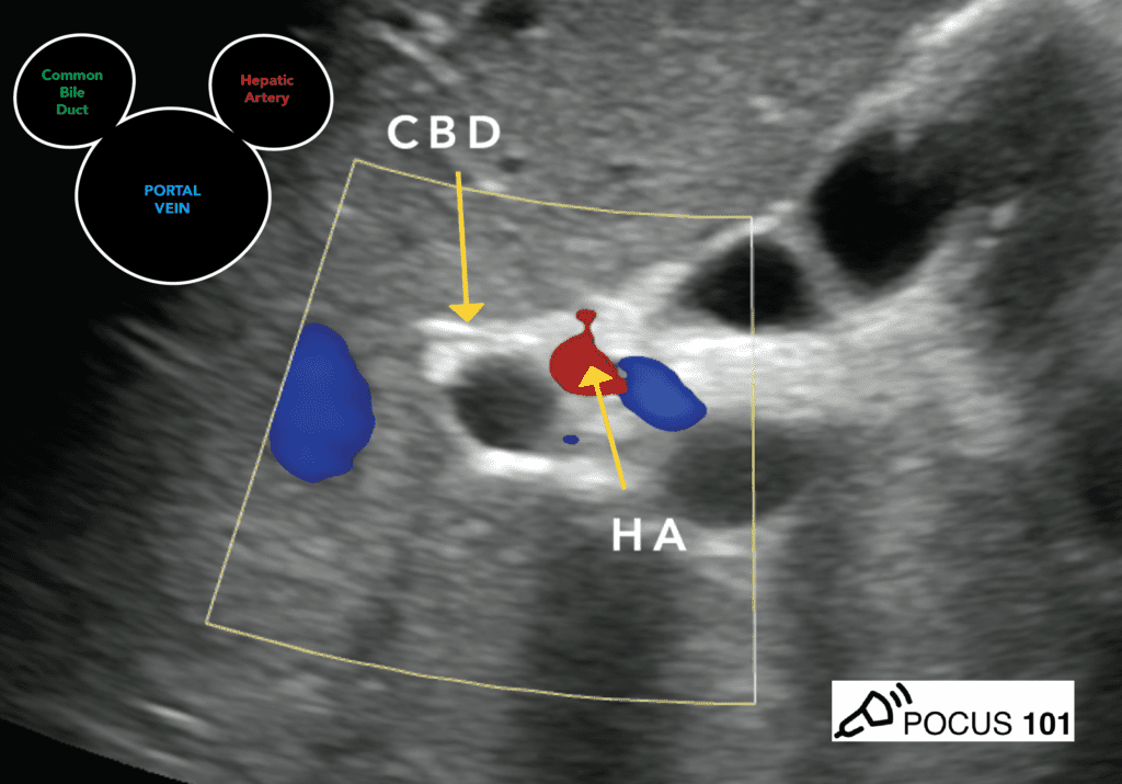
right upper quandrant (RUQ), S. Följ under höger arkus tills Portal Triad = Mickey Mouse Sign : Portal vein (PV), Hepatic artery (HA), Common Bile Duct (CBD)
-
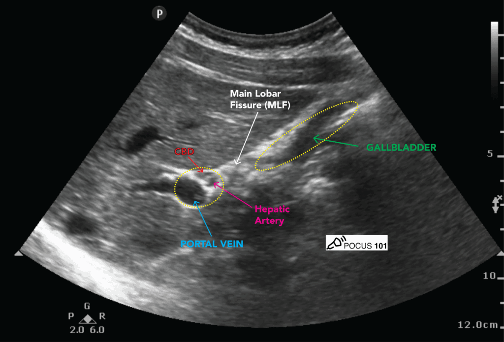
Leta efter Exclamation Point Sign-vyn genom att rotera medsols. Gallblåsehalsen sitter fast vid Main Lobar Fissure (MLF).
-
Leta igenom gallblåsan transversalt efter stenar, genom att Vinkla / Tilt / Fan:
-
Väggtjocklek anteriort, <3mm
-
Kolla CBD
-
Murphy Sign
-
Collins sign
-
“rolling stones”
-
hyperechoic collections with posterior shadowing within the gallbladder lumen
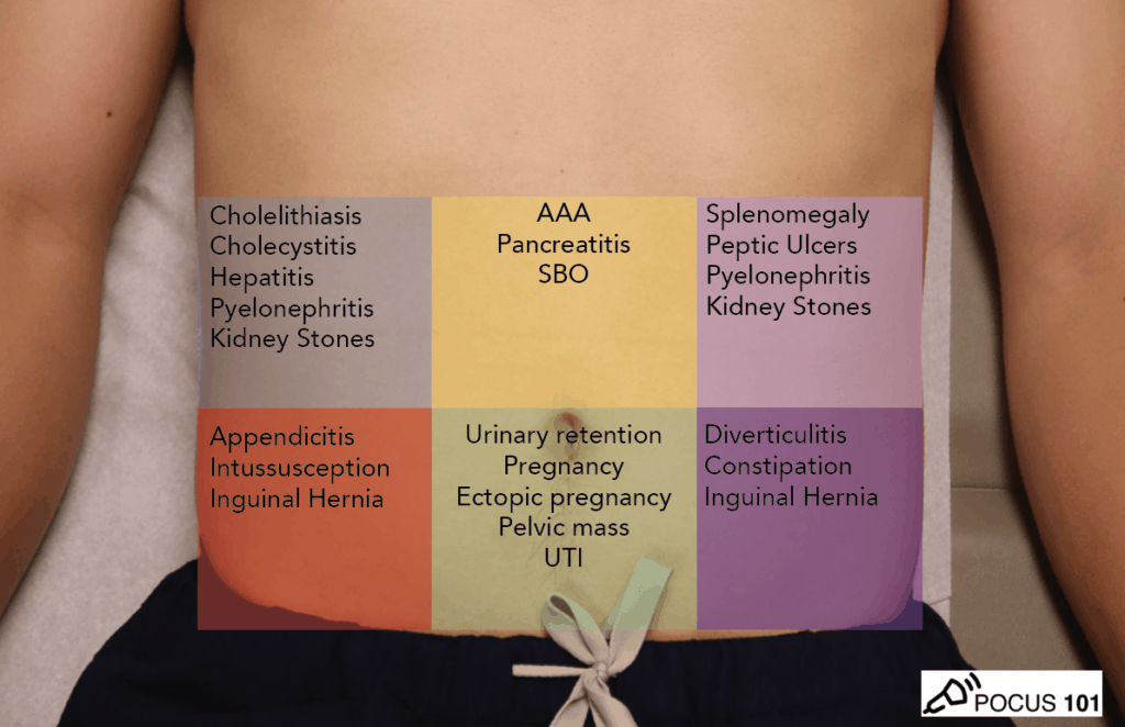
Cholelithiasis refers to gallstone formation anywhere along the biliary tree. Gallstones are present in up to 10% of the population, and only about 1/4 of patients are symptomatic. When symptomatic, the most common finding is biliary colic (post-prandial RUQ pain, especially following a fat-rich meal). Pain may radiate to tip of the right scapula, a finding known as Collin’s sign. Nausea, bloating, belching, heartburn, and flatulence are also common (Bell).
Inga kommentarer:
Skicka en kommentar